Your bones are alive.
Here’s what changes
as you age.
Inside a seemingly solid structure, a repair process never stands still. What happens when its balance begins to shift?
Start with the living structure ↓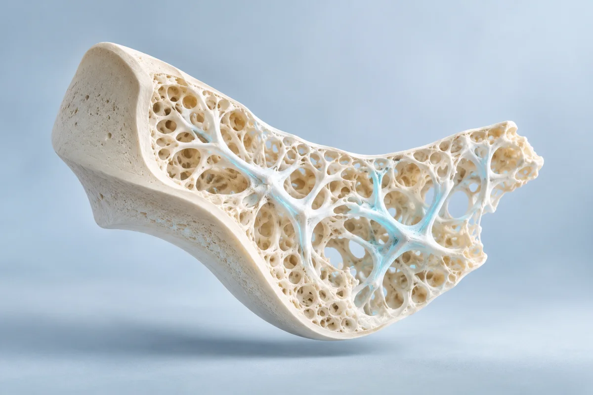
The structure that never stands still.
Quiet on the outside.
Busy within.
Step off a kerb. Carry a shopping bag. Stand up from a low chair. Your skeleton makes these ordinary moments possible. It is easy to imagine it as fixed scaffolding: something built in childhood, then gradually worn down.
But bone is living tissue. Beneath its hard surface, cells maintain a material made from an organic matrix, largely collagen, reinforced with mineral. The combination matters. Mineral contributes stiffness; the collagen-based matrix helps the tissue resist damage. Bone is a composite, rather than a solid piece of chalk. [1]
There is architecture here, too. Cortical bone forms a relatively dense outer layer. Trabecular bone is an internal network of plates and struts, especially prominent near joints and in vertebrae. Their proportions vary around the skeleton. An open network is not automatically a defect; it is part of how bone is organised. [2]
That distinction changes the question we ask about ageing. It is not only “How much bone is left?” It is also “How is the material arranged, and how well is it being maintained?” To understand the answer, start with a surprisingly necessary act: taking a small piece of bone away.

A repair crew,
working in sequence.
Bone does not stay functional by remaining untouched. Small areas can be removed and replaced through bone remodelling. Many sites can be at different stages at the same time: maintenance carried out within a structure that still has to support you. [3]
The cells within the structure
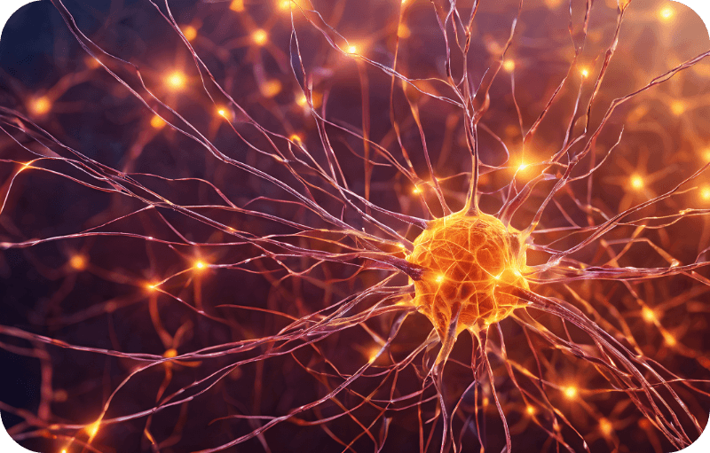
Osteocytes are mature bone cells embedded in the tissue. A network of fine connections helps them communicate and respond to their mechanical environment. Think of them as part of the monitoring system, rather than bricks trapped in a wall. The analogy has limits: these cells are alive, respond to chemical signals and help coordinate other cells. [2]
The key is a sequence, with a handover between the cells that remove tissue and those that rebuild it. Follow one site below to see why those are different jobs. [3]
Life inside the material.
Embedded osteocytes help coordinate responses to loading and local conditions. They are part of a communicating network, rather than passive occupants. This overview starts with sensing; it is not a claim that one cell alone initiates every repair site. [9]
Remove a patch.
Osteoclasts dissolve mineral and break down the underlying matrix, creating a small excavation. Removal is normal maintenance; what matters is how it is regulated and what follows. [1]
Prepare for formation.
Between removal and rebuilding, cells prepare the surface and signals link the two activities. Human biopsy research implicates reversal cells in this handover. This transition is part of the process, rather than an empty pause. [13]
Lay down new matrix.
Osteoblasts deposit osteoid: new organic matrix rich in collagen. The framework comes before its full mineralisation. “New bone” is therefore something that develops and matures, rather than appearing fully finished. [1]
Reinforce the framework.
Mineral is incorporated into the matrix as the tissue matures. Sites eventually return to a resting state. The stages overlap in a living skeleton; this five-part explanation is a teaching aid, not a timed treatment model. [3]
Why a small shortfall can matter
Imagine maintaining a network of narrow footbridges. Replacing worn sections makes sense. Leaving a slightly thinner replacement each time would gradually change what the network can carry. Bone is more complex, but the comparison captures the importance of a repeated process.
A small mismatch can look unremarkable in isolation. Repeated across many sites and over time, it can become a structural problem. This is why the balance between removal and formation matters, as well as the amount of activity. [3]
More activity is not always better
A busy repair crew can still leave work unfinished. In human bone biopsies, researchers found reversal cells with features suggesting arrested activity, associated with reduced bone volume and formation surfaces in postmenopausal osteoporosis. This observational tissue study suggests the handover can matter; it does not establish that every ageing skeleton has the same fault. [13]
Why remove bone at all?
Normal remodelling renews tissue and contributes to mineral regulation. Removal is a necessary part of that maintenance, rather than an error to eliminate. [3]
The diagram and bridge analogy explain a process. They do not model an individual’s bone health.
Small shortfalls.
A different structure.
Ageing can shift the balance so that rebuilding does not fully replace the bone removed. The consequences are visible at a scale much smaller than the outline of a skeleton. In cortical bone, internal spaces can enlarge and the remaining layer can become thinner. Trabecular architecture can also deteriorate, with patterns varying by sex and skeletal site. [4] [14]
Picture the difference between narrowing a supporting beam and losing the connection at its end. Both remove material, but the effect on a structure also depends on where the change occurs. That is why the phrase “bone loss” is useful yet incomplete. It describes a reduction without showing its geometry.
Changes within the outer shell.
In a study of 122 women aged 27–98, researchers combined high-resolution wrist imaging with tissue analyses to examine cortical and trabecular bone. Their findings highlighted how increasing cortical porosity contributes to age-related structural deterioration. This was a cross-sectional comparison across ages, not a record of each woman’s skeleton changing over decades. The distinction matters: a clear pattern is evidence, but it is not an individual forecast. [4]
Why menopause can accelerate the change
Age and menopause overlap, but they are not interchangeable explanations. Oestrogen helps regulate the signals controlling bone turnover. Its decline around menopause can shift the system towards greater resorption. A small human mechanistic study found differences in RANKL expression, a signal involved in osteoclast formation, after menopause. That helps explain a pathway; it does not mean one signal accounts for every woman’s bone loss. [5]
A transition, rather than a single birthday
The longitudinal SWAN study followed 1,902 women through the menopause transition. Bone-density loss accelerated in late perimenopause and remained substantial in the early postmenopausal years. Unlike comparing younger and older groups once, repeated measurements can show when changes gather pace. The trajectory still varies between people. In this analysis, the average follow-up was 3.9 years, with measurements at up to six annual visits. The study maps a change in pace around a transition; it does not supply a fixed amount that every woman will lose. [6]
Men also experience skeletal ageing. A population-based study comparing wrist microstructure in 324 women and 278 men found different age-associated patterns between the sexes. This is another reason to avoid one universal picture of an “old bone”. Life stage, sex and skeletal site help shape the story; a birthday alone cannot describe it. [14]
Density is useful.
It is not the whole structure.
A DXA scan uses X-rays to estimate areal bone mineral density: mineral content relative to the projected area of the bone being measured. It is an established part of osteoporosis assessment. But a two-dimensional density measure does not describe every strut, pore, material property or aspect of bone shape. [8]
This is not a reason to dismiss the test. It is a reason to interpret it in context. A useful photograph of a building does not become useless because it cannot show the condition of every joint. Equally, it should not be asked to answer questions outside its view.
Research supports that distinction. In the BoMIC prospective study, 7,254 older women and men were assessed with high-resolution peripheral CT and followed for fractures. Measures of microarchitecture and estimated strength added information beyond hip DXA density and FRAX risk estimates. These research tools explore structure in detail; the study does not mean everyone needs a specialist CT scan. [7]

Amount, arrangement and the load.
Mineral in a projected area.
DXA divides the measured mineral content by the projected bone area. This gives a useful density measure, expressed in grams per square centimetre. It does not directly map each internal connection. [8]
Withstand the force it faces.
Architecture and geometry influence how a load is distributed; material properties influence how tissue responds. A pore or a missing connection changes the structure in a different way from uniformly reducing its size. Bone amount still matters, alongside its arrangement. [7] [2]
A density result helps describe bone.
Fracture risk also belongs to the person.
Previous fractures, age, some medicines, health conditions and the likelihood of falling can change the clinical picture. A clinician combines relevant information rather than treating one number as a guarantee. [15]
Bone listens to
the loads it carries.
The skeleton is a structure with sensors. Mechanical loading can be translated into cellular signals that influence bone formation. In animal experiments, loading changes osteocyte signalling. They explain plausibility; they do not tell us how much a particular person’s bone density will change after an exercise programme. [9]
For that, we need human studies—and we need to read their details. A result obtained with selected participants, a particular programme and professional supervision is not a promise attached to the word “exercise”.
What one carefully supervised trial found.
The LIFTMOR randomised trial enrolled 101 postmenopausal women with low bone mass, with an average age of about 65. It compared eight months of twice-weekly, 30-minute supervised high-intensity resistance and impact training with a home-based low-intensity programme. Participants were screened before enrolment. [10]
Reported group means after eight months; BMD = bone mineral density. These are study results, not predicted personal outcomes. The trial reported variation around each mean. [10]
A meaningful finding, with boundaries
Physical function also improved, but the trial did not establish fracture reduction. The density changes cannot be converted into a claimed reduction in fracture risk. One minor lower-back spasm was reported. These supervised results do not establish the safety of unsupervised heavy lifting. [10]
What the result makes possible
The finding makes a useful question possible: what challenge fits this person? A trial can inform that conversation without supplying a universal workout.
A more useful next step
than a promise.
It is tempting to reduce a living system to a single fix. Bone biology points towards a better question: which part of the picture needs attention?
Keep an appropriate challenge.
Guidance supports weight-bearing activity and resistance exercise adapted to ability. Balance and strength work also help address falls. The useful goal is regular, suitable activity; a study of supervised training is not a prescription to copy its heaviest exercises. [11]
Attend to the materials.
Adequate nutrition, including calcium and vitamin D, belongs in bone care. Calcium-rich foods help meet intake needs; vitamin D helps calcium absorption. Supplementation may be appropriate when intake or status is inadequate. More is not automatically better, and correcting a shortfall is different from proving that a product prevents fractures. [12]
Think beyond the skeleton.
Muscle function, balance and the environment influence the circumstances of a fall. Bone care and falls prevention address related parts of the problem. This wider view makes everyday function a useful part of the conversation. [11]
Consider two people who both want to “look after their bones”. One may need help building a consistent routine. Another may have had a fracture after a minor fall and need a clinical assessment. The same slogan does not answer both situations. The useful action comes from identifying the problem, rather than choosing the most ambitious claim.
That is a helpful way to read longevity research, too. Ask what changed, in whom, over how long, and under what conditions. A cell signal, a density measurement, a physical-function test and a fracture are different outcomes. Each can teach us something, provided we keep its meaning intact.
Still living.
Still responding.
Your skeleton is not simply a framework slowly running out of material. It is tissue being maintained, with a balance between removal and rebuilding and an architecture that helps distribute load.
Three ideas make the subject easier to understand: repair is a sequence, small imbalances can accumulate, and density is one useful view of a larger structure. None makes ageing irrelevant. Together, they show why bone health deserves a more thoughtful conversation than “strong” or “weak”.
The most useful long-term perspective is neither resignation nor a promise of reversal. It is attention to a living system: the demands it faces, the resources it needs and the evidence that can guide the next decision.
Sources & further reading.
Primary human studies support the age, menopause, architecture and exercise examples. Physiology reviews explain the process; clinical guidance informs the practical context. The loading experiment is animal research. These evidence types answer different questions.
- NIAMS: What Is Bone? ↗Authoritative physiologyLiving tissue, composition, cell roles and remodelling.
- Florencio-Silva et al. (2015): bone tissue and its cells ↗Physiology reviewMatrix, cortical/trabecular structure and osteocyte communication.
- Raggatt & Partridge (2010): mechanisms of remodelling ↗Physiology reviewActivation, resorption, reversal, formation and termination.
- Zebaze et al. (2010): cortical remodelling and porosity ↗Primary human study / cross-sectionalAge comparisons in women; imaging and tissue analysis. Author-institution abstract.
- Eghbali-Fatourechi et al. (2003): RANKL after menopause ↗Primary human mechanistic studySmall, nonrandomised marrow-cell comparison; not a treatment recommendation.
- Finkelstein et al. (2008): SWAN menopause-transition study ↗Primary human study / longitudinal cohortRepeated bone-density measurements in 1,902 women.
- Samelson et al. (2019): BoMIC microarchitecture study ↗Primary human study / prospective cohortsFracture prediction beyond DXA and FRAX; not a routine CT-screening recommendation.
- Lewiecki (2024): osteoporosis clinical evaluation ↗Clinical measurement reference / EndotextDXA measurement. Public overview: NIAMS bone-density tests. NIAMS overview of bone-density tests ↗
- Robling et al. (2008): loading and osteocyte signalling ↗Primary animal experimentMechanistic evidence; not a human exercise outcome.
- Watson et al. (2018): LIFTMOR randomised trial ↗Primary human randomised trialScreened postmenopausal women; supervised training; density and function endpoints. Published correction to table headings and calcium-intake sign ↗
- NOGG: non-pharmacological management ↗Clinical guidelineActivity, nutrition and falls care, tailored to the person.
- Healthy Bones Australia: calcium and bone health ↗Australian consumer guidanceFood, supplementation when needed and the role of vitamin D. Healthy Bones Australia: vitamin D and bone health ↗
- Andersen et al. (2013): the reversal-cell handover ↗Primary human biopsy studyObservational tissue evidence. Author-institution abstract.
- Khosla et al. (2006): age, sex and microstructure ↗Primary human study / cross-sectionalWrist microstructure in women and men; patterns are not individual trajectories.
- NOGG: fracture-risk assessment ↗Clinical guidelineDensity interpreted alongside clinical history and other risk factors.
This is general educational context; individual fracture-risk, exercise and treatment decisions need personal clinical assessment. Illustrations simplify structure and do not show measured outcomes.